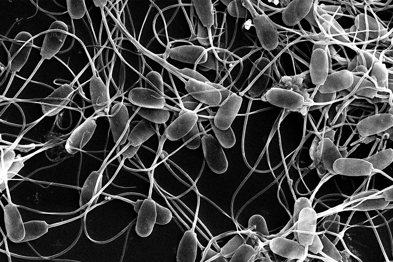
In a previous blog post, we talked about the parts of the spermatozoon and spermatogenesis . Today we will focus on the last phase of this process, that of differentiation or spermiogenesis, to explain how this particular cell is formed in more detail.
During spermiogenesis the initial spermatids differentiate into spermatozoa, after undergoing a series of structural and biochemical changes:
- Condensation of the nucleus: the nucleus of the spermatids is compacted and acquires an oval and pyriform shape. The DNA is also compacted by the replacement of histones with protamines, which are proteins that will stabilize the nucleus until fertilization.
- Acrosome formation: The acrosome is a cap covering the head’s apical area. It contains hydrolytic enzymes which, after the acrosomal reaction, will allow the sperm to pass through the zona pellucida and fertilize the egg.
- Reduction of the cytoplasm: most of the spermatozoa cytoplasm is eliminated by its displacement towards the end piece of the flagellum, originating the so-called cytoplasmic drop, which must be eliminated at the end of the sperm maturation process in the tail of the epididymis. The presence of spermatozoa with cytoplasmic droplet in an ejaculate is a sign of immaturity and is classified as a secondary malformation.
- Development of the flagellar movement system: the centrosome of the spermatozoon (skeletal system of the cell) moves towards the opposite side where the acrosome has formed. Thus, on the one hand, the neck of the spermatozoon is assembled, which allows the anchoring of the flagellum to the head and, on the other hand, the axoneme of the flagellum is formed. Around the axoneme, the so-called external dense fibers are formed, which will extend along the main part of the flagellum, and which are responsible for the bending movements of the flagellum.
- Formation of the mitochondrial helix: mitochondria migrate from the head and are positioned in the proximal part of the flagellum, helically surrounding the axoneme and giving rise to the middle piece of the flagellum. These mitochondria are responsible for providing the energy that activates flagellar movement.

Figure 1. Representative scheme of the spermiogenesis process.

Figure 2. Representative scheme of the spermiogenesis process. Modified from Essentials of human anatomy & physiology (Marieb, 2006). Source: Sara Miguel Jiménez doctoral thesis (2023).
When the spermatozoa complete spermiogenesis they are not yet functional, but must undergo post-testicular maturation. First, in the epididymis, they will finish maturing and acquire the ability to move, and in the reproductive tract of the female, they will complete the process of sperm capacitation, which allows them to finally fertilize the egg. About this, we delved more in previous entries of this blog, which you can consult in: “The journey of the sperm towards fertilization“.
Bibliography
Text adapted from the doctoral thesis “Study of the possible antioxidant, antiapoptotic and chemotactic action of melatonin on ovine spermatozoa”. Sara Miguel Jiménez. University of Zaragoza (2023).
The sperm journey to fertilisation
Marieb (2006). Essentials of human anatomy & physiology. San Francisco, Pearson/Benjamin Cummings.
Senger (2003). Pathways to pregnancy and parturition, Current Conceptions, Inc., 1615 NE Eastgate Blvd.
Lindeman (1996). Functional significance of the outer dense fibers of mammalian sperm examined by computer simulations with the geometric clutch model.Cell Motil Cytoskeleton 34(4): 258-270.
Turner 2003. Tales from the tail: what do we really know about sperm motility? J Androl 24(6): 790-803.
Reproductive physiology of the boar: Spermatogenesis, SUIS Nº 75 March 2011
Boar Reproductive Physiology: Sperm Physiology and Sperm Production, SUIS No. 76 March 2011