In this blog post we will give a series of basic notions about the anatomy and reproductive physiology of the boar. It will help us to understand how sperm production works and how it is regulated at the hormonal level.
Anatomy
The reproductive tract of the boar consists of different structures:
Testicular and epididymis anatomy
We will detail a little more these two parts since in them sperm are produced and mature, so their knowledge will help us to understand possible alterations of sperm quality that we see in the laboratory.
The testes are the gonads, whose two main functions are: cytogenic function (sperm production) and endocrine function (production of male sex hormones). The scrotum has a protective and thermoregulation function maintaining a temperature of 3-4ºC lower than that of the rest of the body. Thermoregulation occurs thanks to the Dartos tunic under the skin layer. It is a fibroelastic tissue composed of muscle fibers able to contract and bring the testicles closer or away from the abdominal wall and the pampiniform plexus, allowing the thermal exchange that occurs in the testicles and keep at 32-34ºC. This function is of great importance as alterations in the temperature inside the scrotal pouch will lead to problems during spermatogenesis and during sperm maturation in the epididymis. A common pathology that can affect the exchange of temperature produced in the pampiniform plexus, is the varicocele.

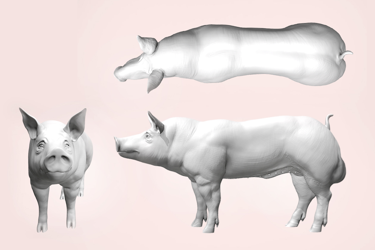
Picture 1: Boar reproductive tract
-
-
- Prepuce
- Preputial sac
- Penis
- Bladder
- Spermatic blood vessels
- Ureter
- Seminal vesicles
- Prostate gland
- Bulbo urethal gland
- Vas deferens
- Epididymis (Tail)
- Testicle
- Scroturn
- Epididymis (Head)
-
The testicles are composed of seminiferous tubules, which is the producing unit, these tubules are organized into lobes. Inside the tubules, sperm cells will be formed, these tubules are covered by a seminiferous epithelium formed mainly by:
- Sertoli cells play a fundamental role in testicular development as they nourish and protect germ cells. These cells during fetal development are responsible for sexual differentiation and, as we will see later, in spermatogenesis. The proliferation of these cells occurs in two waves the first over the first month of life and the second at 3-4 months. FSH produced in the pituitary gland has a direct effect on the maturation of these cells. Sertoli cells stop dividing before puberty (regulated by thyroid hormone T3) and their number will determine future sperm production, since without the structural and metabolic support of these cells, the germ cells would not be able to perform spermatogenesis. For this reason, the measurement of testicular size in the pre-pubertal stage is so important when selecting boars for production.
- Leydig cells, or steroididogenic cells, have the ability to synthesize and secrete steroids, enzymes, and peptides. Its function is mainly controlled by LH, although FSH also regulates the functioning of these cells. These cells are located in the interstitium around the blood vessels so their hormonal secretion easily passes into the circulation. These cells provide support and are responsible for the production of testosterone. In contrast to Sertoli cells, more than the size of them, the capacity to produce testosterone is linked to the amount of smooth endoplasmic reticulum present in them. Furthermore, they are responsible for sexual differentiation, testicular growth and the appearance of secondary male sexual characters and androgen-dependent structures and spermatogenesis in the boar.
- Germ cells: the proliferation of these cells during the first weeks after birth is responsible for the formation of seminiferous tubules. The number of germ cells in the testicle increases continuously, with a peak of division at 4-5 months and a stabilization of the population after 7 months. These cells are responsible for carrying out spermatogenesis, through a series of mitotic missions, followed by a series of meiotic divisions ending with the result of a haploid cell.

Picture 2: Seminiferous tubule histological picture
Source: University of Zaragoza
These tubules flow into the rete testis that transports sperm through the efferent ducts, thanks to its ciliated epithelium, until they are deposited in the head of the epididymis. Where the so-called epididymal maturation will begin.

Picture 3: Seminiferous tubule histological picture
Source: University of Zaragoza
Epididymis, is a unique tube contoured on itself, with a length between 50 and 60 meters long, which condenses in 15 to 20 cm, it is attached to the dorsal part of the testicle, it has three parts according to its anatomical position and its function: head, body and tail. Histologically along the duct, we can differentiate four cell types:
- Main or columnar cells, responsible for the transport, secretion, synthesis and absorption of molecules.
- Basal cells, metabolic waste disposal.
- Clear Cells, remove any material or residue present in the lumen.
- Halo cells, belonging to the immune system.
The function of the epididymis is to transport, mature and store sperm. For this purpose along the epididymis are synthesized and secrete proteins. This function is going to be regulated hormonally by androgens and whose function will be to make structural and functional changes in the membrane and structure of sperm. During this maturation process, the sperm acquires the ability to move and fertilize. This maturation occurs mainly in its transit through the head and body of the epididymis. This transport occurs due to the peristaltic movements of the duct. The changes in the sperm cell will not only be morphological, but also biochemical and physiological by interacting with the secretions of the epididymis.
From this phase come the so-called secondary anomalies, characterized by the presence of cytoplasmic droplets either proximal or distal, the latter being considered a milder ripening problem, we can include other anomalies caused during epididymal maturation such as agglutination and the presence of anomalies in acrosomes, although these may also be caused by external factors.
The vas deferens is a tube-shaped structure, surrounded by smooth muscle fibers, which connects the tail of each epididymis with the urethra, its function is to drive the sperm into ejaculation. This structure is part of the spermatic cord.
Set of accessory glands, whose activity depends on the excretion of androgens and are responsible for the production of the non-sperm fraction of the ejaculate (seminal plasma). These glands are:
- Seminal vesicles: they are two glands, joined together, located dorso-lateral to the neck of the bladder, and they flow into the urethra through a proper duct. Its secretion is part of the post-sperm fraction, where the number of sperm is lower. They produce a fructose-rich alkaline secretion that protects the sperm from abrupt changes in pH and gives volume to the ejaculate.
- Prostate: odd and smaller gland in the boar. It secretes a liquid rich in amino acids, citric acid and enzymes. The function of these components is to stimulate the movement of sperm. Along with the secretions of the seminal vesicles, it will form the seminal plasma of the sperm and post-sperm fraction.
- Bulbo-urethral glands or Cowper’s glands, paired glands that are dorsally located to the most caudal portion of the pelvic urethra. They secrete a substance rich in mucin, forming the so-called “tapioca”. Together with part of the prostate secretion, it constitutes the pre-espermatic portion of the ejaculate, whose function is to clean the urethra, therefore it is the first portion of the ejaculate that is usually discarded during extraction, it is rich in components that will promote sperm motility in the newly ejaculated sperm.
Urethra, is a tubular structure that begins in the bladder, which conducts the sperm from inside the abdomen through the inguinal canal to the external urethral orifice located at the vertex of the penis, being able to differentiate two parts, the pelvic urethra and the penile urethra. The urethra leads the semen and urine to the outside, therefore it is important to discard the first fraction of the ejaculate, to prevent cross contamination.
Penis, the boar has a fibrous penis. It is the external sexual organ that allows the mating, and the urethra flows through its interior to the glans. The penis is formed by the body of the penis and the glans, both covered by the foreskin. The penis of the boar has a sigmoid inflection that, when extended, produces the exteriorization of the organ. In the foreskin we can find two blind sacs, called preputial sacs where urine and remains of previous ejaculations accumulate, in addition in the foreskin are present a series of preputial hairs. These two structures can become a great source of contamination, so it is advisable to empty the preputial sacs, trim the hairs frequently and clean the foreskin before each extraction.
With this blog post we want to give a brief reminder of the anatomy of the porcine stallion. In the next post we will talk about the physiology and hormonal control of spermatogenesis. Both entries will serve as an introduction in the production part of seminal doses. Once we know the structures and how sperm cells are produced, we will be able to influence and deepen more in the different functions and tips to optimize the production to the maximum in the boar stud.
BIBLIOGRAPHY
Bonet, S., Garcia, E., Sepúlveda, L. 2013. The Boar Reproductive System. In: Bonet S., Casas I., Holt W., Yeste M. (eds) Boar Reproduction. Springer, Berlin, Heidelberg.
Úbeda, J.L., Dahmani, Y., Ausejo, R. (Marzo, 2011). Fisiología Reproductiva del Verraco. Suis, 75: 32-41.
Grandía Ansó J, Ansó T, Ausejo R, Suárez A, Mitjana O, Falceto MV. 2020. Ecografia testicular aplicada a la evaluación seminal en verracos. ANAPORC, Febrero 2020, 24-28.
Romeo R.D., Richardson H.N., Sisk C.L. 2002. Puberty and the maturation of the male brain and sexual behavior: recasting a behavioral potential. Neuroscience & Biobehavioral Reviews. 26, (3): 381-391.
França L.R., Avelar G.F., Almeida F.F. 2005. Spermatogenesis and sperm transit through the epididymis in mammals with emphasis on pigs. Theriogenology, 63(2), 300-318.
https://www.thepigsite.com/genetics-and-reproduction/insemination/anatomy-and-physiology-1
https://www.3tres3.com/articulos/anatomia-y-fisiologia-del-verraco_4025/