In this blog post we are going to talk about the anatomy and physiology of the sow’s reproductive system, which has a large length and covers from the vulva to the ovaries, all structures are suspended by the wide ligament. The reproductive system consists of the following parts:
- Vulva: external genital part, it is externalized with two lips, at the bottom of them is the clitoris. The vulva is highly vascularized, changing its morphological appearance during the heat, being much more inflamed and edematous. This feature is much more marked in gilts, and it serves as one more tool to detect if a sow is in heat.
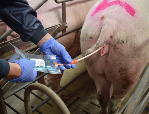
- Vestibule and Vagina: next to the vulva we find the vaginal vestibule and the vagina, with a variable joint length between 30-40 cm, in whose caudo-ventral part is the urinary meatus. This structure forces the catheter to be inserted with an angle of about 30-45º, to the urinary tract. The vagina in its cranial part connects with the cervix, has an immunoprotective function with the presence of IgA.

Picture 1: Reproductive system
- Cervix: it is a complex structure and because of this, desingning probes able to cross it without causing injury to it and being able to carry out the technique of post-cervical insemination acquires great importance. This structure is about 2-3 cm in diameter and about 15-20 cm in length and forms the gateway to the uterus. It is formed by cervical folds or tuberosities, these folds change consistency due to hormonal influence. Remaining closed in the presence of progesterone and staying dilated during estrus. It also has a secretory function of mucous substance, which serves as lubricant at the time of mounting and protective function during pregnancy.

Picture 2: Cervical rings
- Uterus: it is formed by the body of the uterus (4-5 cm) where it bifurcates into two uterine horns. The horns have a variable length between individuals due to various factors including race, number of births and age. They have a tortuous shape and connect the body of the uterus with the oviducts. This is where the embryos will be implanted. In fact, the maximum number of piglets per litter depends greatly on the uterine capacity. The uterus has a wall composed of three layers, the endometrium, myometrium and serosa, presents uterine glands along the entire uterine tract essential in pregnancy.
- Oviduct: they are thin and long-distance ducts of about 25 cm that join the uterus to the ovaries, which are divided into three regions: infundibulum, blister, and isthmus. The part closest to the ovary, the infundibulum, surrounds it forming what is known as the ovarian pouch, while the isthmus is attached to the uterus by the uterus junction, in this area where the sperm remain viable and with fecundating capacity about 24 hours, forming the sperm reservoir.
- Ovaries: they are the female gonads, and they have two main functions, the first is the production of oocytes and the second is the synthesis of reproductive hormones. Their shape is lobed, they are not physically connected to the rest of the reproductive system, they are attached to the rest by the broad ligament of the uterus.
Once the anatomy of the reproductive system has been explained, we can begin to explain the physiology to see how the structures described above perform their functions.
Puberty is the moment when the female begins her reproductive activity and therefore the first ovulations appear with the first symptoms of heat. In most females puberty is reached between 180-220 days of life and 120 kg of weight. But it depends to a large extent on different internal factors, such as the breed or the genotype of the sow or external factors such as the time of year, nutrition, transport and the correct adaptation to the farm, the housing management (in groups or in individual boxes) and exposure to the male. These latter external factors can be optimized to anticipate the onset of puberty and increase herd performance.
Once the female begins her reproductive activity, her sexual cycle is repeated every 18-24 days continuously, this is because the sow is a continuous polyestrica specie. This cycle is only interrupted by pregnancy or lactation. This process is controlled by hormones that are synthesized in the anterior pituitary gland (FSH, LH, prolactin), as well as intrafollicular factors (steroid hormones, peptide hormones, prostaglandins, and growth factors) (Figure 1).

Figure 1: Hypothalamic-hypophysary-ovarian axis
The sexual cycle of 21 days (Figure 2), can be divided into two phases, luteal phase and follicular phase, within these two phases we find four states.
Follicular phase (4-6 days)
- Proestrus
- Estrus

Figure 2: Hormone levels in the blood throughout the estrus cycle (Adapted from Laing et al, 1991)
Luteal phase (14-16 days)
- Metaoestrous
- Diestrus
From day 1 to 3 of the sexual cycle (estrus), during this phase in which the high concentration of estrogens and inhibin that inhibit the secretion of FSH predominate, terminal follicular growth occurs. We can find ovaries with prominent and turgid 8-12 mm follicles with fine vascular reticulation on the surface. Transparent walls and reveals a straw colored fluid. On many occasions we can identify an area that indicates the future ovulation point (stigma or avascular papilla). Albicans bodies may also appear as remains of the corpus luteum from the previous cycle.
Days 3-4 of the cycle (metaestrus): ovaries with follicles about to ovulate and newly ovulated rubrum bodies that are organizing the clot that has remained after the rupture of the follicles. The rubrum or hemorrhagic bodies have a collapsed appearance, a conical shape and a dark red color, and they show the point through which the ovum has left the follicle.

Source: M.V. Falceto

Source: M.V. Falceto

Figure 3: Reproductive cycle and follicular and luteal dynamics diagram of the sow (adapted from Hafez, 1987; Mc Donald, 1991)
Days 5 to 14 of the sexual cycle (luteal progressive phase), this phase is characterized by the secretion of progesterone by luteal bodies, whose functions are to induce the proliferation of the endometrium (embryonic survival) and block the development of follicles (blocking FSH and LH).
- During the first 48 hours of the luteal phase, we observed rubrum/luteal bodies that are red. Knowing the number of rubrum bodies present in the two ovaries we determined the ovulation rate.
- Subsequently, luteal bodies of 8-11 mm are observed that have a fleshy appearance. The surface is vascularized and the ovulation point disappears.
- From the 8th to the 14th we observe corpus luteum of 10-15 mm. On day 10 the maximum ovarian weight and the maximum progesterone values are reached in the hormonal determination. At this time, if the female is pregnant, she will receive an embryonic signal in the form of estrogens secreted by the embryos that will prevent the regression of the corpus luteum. For this to happen, a minimum of 4 embryos are needed to occupy both uterine horns on 70% of their surface.

Source: M.V. Falceto
")
Source: M.V. Falceto
Days 15-16 of the sexual cycle (luteal regressive phase): ovaries with pale pink corpus without vascularization. During this phase the ovary presents its minimum size and luteolysis is evident, producing a rapid decrease of progesterone to basal levels and an increase of prostaglandins. Follicles smaller than 4mm will suffer atresia, while those larger than 4mm will continue their growth.

Source: M.V. Falceto
Days 17 to 21 of the sexual cycle (proestrus): it is the moment when the ovary prepares for the next cycle, the selection and growth of the dominant follicles will take place (days 17-18), the pituitary secretion of FSH increases, with its subsequent fall, and followed by an increase in the concentration of estrogens that cause a preovulatory LH peak. In this phase, large ovaries with hyperemia appear where 10-25 follicles 8 mm are found, accompanying several 3-5 mm albicans with a yellowish or white cream color, which will gradually disappear until they become a scar in fibrous tissue of the ovary.

Source: M.V. Falceto
We hope with this information have supplemented in a certain way your knowledge about “what happens” inside the sow during the estrus cycle, and to better understand the reason for the different actions that are carried on the day to day in the farm. In next blog posts we will focus on a topic closely related to this, such as the guidelines for a correct induction of heat in the sow and the insemination technique.
Don’t miss it!
BIBLIOGRAPHY
Dyce KM. 2009.Textbook of Veterinary Anatomy, Ed St. Louis: Saunders/Elsevier
Gil, M.A., Cuello, C., Parrilla, I. (2009) Fisiología del tracto genital de la cerda y el verraco. Cuaderno de Reproducción Anaporc. 55: 24-31.
Falceto, M.V. (1992) Aportaciones al estudio de la estacionalidad reproductiva en la cerda. University of Zaragoza
Falceto, M.V., Duque, C., Alfonso, J., Ciudad, M.J., Espinosa, E. (2004) Variaciones Fisiológicas de la funcionalidad ovárica en la cerda. Porci. 82: 11-32.
Hafez ESA. Reproduction in farm animals. Lea & Febiger, 2000.
Mc Donald LE, Pineda MH. Veterinary Endocrinology and Reproduction. Lea& Febiger, 1991.
Laing JA, Brinley WJ, Wagner WC. Fertilidad e infertilidad en la práctica veterinaria. Interamericana-Mc Graw-Hill, 1991.