Ultrasound is a diagnostic imaging technique based on ultrasound, which allows a quick and non-invasive clinical study. Ultrasounds are mechanical vibrations with frequencies higher than those audible to humans and have the following characteristics:
- Wavelength corresponds to the distance between two points in the wave trajectory.
- Frequency, which determines the depth that the ultrasound reaches. It should be taken into account that the higher the frequency, the lower the penetration in the tissues and the higher the quality of the image.
- Amplitude, which indicates the intensity of the sound, and is expressed as greater or lesser intensity of the target.
- The velocity of propagation represents the space that the wave travels per unit of time.
Ultrasound scanners consist of three parts, the transducer or probe, the processing unit, and the screen. The transducer or probe consists of crystals that make movements to emit and receive waves. The gel that is placed on the probe to make contact with the animal to be examined is used to favor the transmission of ultrasound.
With ultrasound, different ultrasound wave amplitudes are measured, and it is manifested with intensities of white, black, and gray.
- The whiter the image (hyperechogenic), the greater the amplitude, and corresponds to bone and air (they reflect most of the ultrasound).
- The images that appear as a grayscale (hypoechogenic) are those perceived by soft tissues that partially reflect ultrasound.
- The blacker the image (anechogenic), the lower the amplitude, and it corresponds to the liquid (they do not reflect the waves).
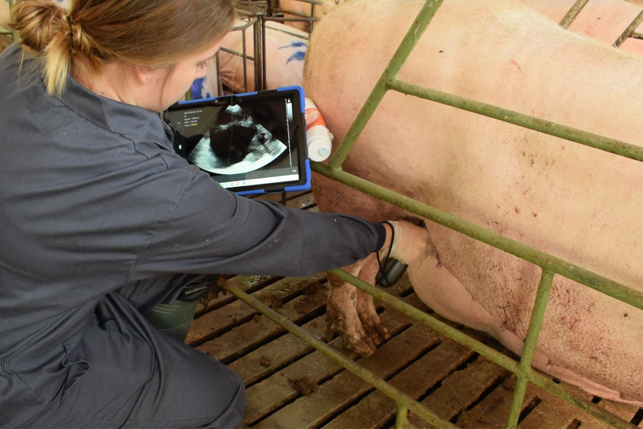
In the swine industry, ultrasound is usually used to confirm pregnancy in sows, to study the reproductive tract (mainly in replacement sows) to assess their puberty status, to detect possible pathologies, or to measure backfat thickness. Since the structures to be studied are different and are at different depths, the probes used will be different. Normally, ultrasound scans for gestation diagnosis are usually performed between 21 and 23 days after insemination, and, in positive sows, black vesicles are seen, as shown in the image below.

Ultrasound at different days of gestation. Source: 3tres3.com: José Luis Guzmán Guerrero (University of Huelva)
To realize the pregnancy control ultrasound, gel should be placed on the probe and look for the area of the sow’s inguinal region, which is close to the hind limb. It is a globose area on which we should place the transducer and look for the presence of these black vesicles to confirm pregnancy.
In nulliparous sows, it may be necessary to place the probe more towards the front area (more cranial) to locate these vesicles, and this placement may vary depending on the number of days of gestation of the sow.
As for other uses of swine ultrasound besides pregnancy confirmation ultrasound, it is worth mentioning the ultrasound study of the genital apparatus of sows at the time of puberty. Through ultrasonography, the uterus and ovaries of nulliparous sows can be evaluated to check if the ultrasound findings are as expected. The typical structures that would appear in such ultrasounds are follicles between 2 and 5 mm in diameter without corpora lutea.
Another application would be the determination of the ovarian cycle by measuring the follicles. In the moments close to ovulation, the pre-ovulatory follicles change shape and can appear simultaneously as the corpora hemorrhagicae. In short, ultrasound makes it easier to know the follicular evolution and to check if the artificial insemination protocol is optimal according to the moment of ovulation, although it is a technique that requires specific training.
Finally, pathological situations such as single or multiple ovarian cysts can also be diagnosed. Fluids can be observed in the uterus, which when they are not associated with pregnancy, semen, or with the sow being in estrus, will be considered a pathological indicator.
Therefore, in the presence of reproductive failure, ultrasound can be a technique that, together with other diagnostic methods, can detect a problem and reverse the cause.